ABSTRACT
The increased reports of microcephaly in Brazil that began in October 2015 have alarmed the public health community. Measuring that increase is dependent upon the establishment of a base occurrence of this birth defect prior to the arrival of Zika viral illnesses. The Brazilian government reported an average of 156 cases per year from 2010 through 2014. The accuracy of that statistic is in question. It is possible to estimate the number of pre-Zika microcephaly cases in Brazil by examining the incidence of conditions known to cause microcephaly. By using only four of the known causes, the estimated case load exceeds 4,600 annually. This calls into question the historic number of reported cases as well as the provenance of the confirmed microcephaly cases in Brazil since October.
INTRODUCTION
In October 2015, the Brazilian Ministry of Health notified the World Health Organization that it was receiving reports of an unusual number of cases of microcephaly in newborns. The medical community suspected a link between prenatal Zika viral illnesses in expectant mothers and microcephaly in their fetuses and newborns. A reporting system was instituted and established criteria defining microcephaly for reporting purposes.
The Ministry issues weekly reports on microcephaly cases. A recent report accessed covers the period November 8, 2015, through May 28, 2016 and the Ministry has received 7,723 reports of microcephaly. [1] It has investigated about 59% of those reports. A total of 1,489 cases have been confirmed as microcephaly caused by an infection. 3,072 cases have been discarded as normal, or microcephaly not due to an infection. The remainder of the reports continue under investigation.
The United States Centers for Disease Control and Prevention (CDC) accepted that a maternal Zika viral illness is a cause of fetal microcephaly on April 13, 2016. [2] The World Health Organization (WHO) also agrees, stating “Based on a growing body of preliminary research, there is scientific consensus that Zika virus is a cause of microcephaly …” [3]
In the pre-Zika era, the most common medical definition for microcephaly used the circumference of the infant’s skull at 24 hours of age compared to an authoritative growth chart. Both the CDC [4] and the WHO [5] have produced these charts, though they differ. [6]
When the Brazilians began tracking microcephaly that could be related to a Zika viral illness, they established a fixed head circumference as the definition. In October, 2015, the definition was a skull measuring less than 33 cm. In December, the Ministry changed that to a measurement less than 32 cm. In March, 2016, the WHO definition was accepted, “<31.9 cm and <31.5 cm for full-term male and female babies respectively”. [7]
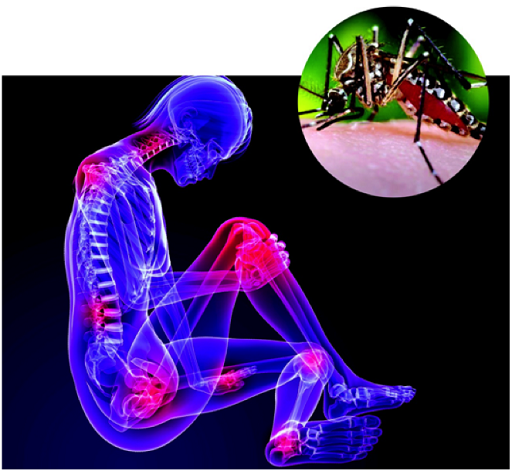
Microcephaly in a fetus is often accompanied by other central nervous system defects or developmental defects. The most severe result in fetal death or the death of the newborn in the first 24 hours of life. Co-morbid conditions and developmental delays after birth can range from severe to non-existent. Microcephaly can also appear in a normal child after birth, usually due to an infection such as chikungunya. [8]
The 7,723 reports in Brazil represent an extraordinary increase in such reports for the nation. In the five year period 2010 to 2014 the nation reported an average of 156 cases per year. [9] That data has been questioned by researchers. As an example, in the period 2006 to 2010, the state of Texas averaged 372 cases yearly in 323,000 annual live births. [10]
METHODOLOGY
Searches for pertinent research were conducted using Google Scholar and Pub Med. The initial search term was “incidence rate” “microcephaly”. This resulted in 1,140 returns in Google Scholar. In addition, the references and citations from pertinent papers were searched manually for additional studies and data sources.
To narrow the focus, the search criteria were changed to reflect the general categories that are known to result in microcephaly, genetic defects, maternal health and behaviors and the infectious diseases cited by the Brazilian Ministry of Health, syphilis, toxoplasmosis, rubeola, cytomegalovirus and herpes. [1] The searches followed the format “incidence rate” “microcephaly”“xxxxxxx”, with the third search term varying.
When data relating to the incidence of microcephaly due to a particular condition or infection was found, additional searches were conducted to determine if there was data specific to Brazil on the same topic.
Four topics illustrative of the known causes of microcephaly were selected for inclusion in this work, Down’s syndrome, Fetal Alcohol Syndrome, congenital toxoplasmosis infections and congenital cytomegalovirus infections. These topics selected were chosen for two reasons. The four topics provided the greatest number of studies and widest availability of data and also provided the widest availability of data from Brazil.
The number of live births in Brazil for 2011 through 2015 was obtained. [11] The incidence of birth defects in Brazil has been studied. Oliveira, in 2011, found that 2.8% of live births had one or more defects. [12] Costa, in 2006, in a large study in Rio de Janeiro, found a prevalence of 1.7%. [13] That study notes that higher prevalences had been reported in the past, likely due to the use of an existing database rather than a hospital-based study.
The greatest risk for error in this work is the varying definitions of microcephaly in use over the last several decades. As noted, Brazil has used three different measurements of head circumference as part of their definition of microcephaly in the last nine months. The growth charts from the CDC and from WHO differ because of the ethnicity and socioeconomic status of the subjects used in each analysis.
Many studies note that the incidence of medical conditions may vary by region in Brazil. That is true for the reports of microcephaly which may be related to Zika, where over 50% of all reports originate in three states, Bahia, Paraíba and Pernambuco.
Another likely source of error, under reporting of microcephaly, appear possible in the literature. Many studies that were read referred to CNS defects or to conditions in which microcephaly could be a subset.
The literature searches were performed in English. It is possible that studies that were solely published in other languages, and in Portuguese in particular, could contain relevant data.
RESULTS
Down’s syndrome is a common genetic disorder. The WHO suggests that the incidence of this condition is between 1 in 1,100 to 1 in 1000 live births worldwide. [14] Costa’s 2006 study found an incidence of Down’s of 6.39 per 10,000 in the Rio de Janeiro region of Brazil. Microcephaly is associated with Down’s syndrome (DS) extensively in the literature.
The incidence of that condition in the DS population is less well documented. One study documented six cases of microcephaly out of 16 patients, 37.5%. [15] Another study found over 60% of 295 patients with DS exhibited microcephaly. [16] A study in SE Brazil of 62 Down’s children found 67% had microcephaly. (17)
Available data was used to estimate the number of Down’s syndrome births in Brazil with microcephaly. The incidence in the 2006 Costa study of 6.39/10,000 was used to estimate total DS births and the number of microcephaly cases in those numbers was calculated using a rate of 60%. That resulted in an estimated 1,133 cases in 2015.
Alcohol consumption during pregnancy is linked to a variety of fetal defects including microcephaly. A study in 1999 estimated the incidence of Fetal Alcohol Syndrome (FAS) in Brazil as 1 in 1,000 births. [18] A study of Native Americans in the United States published in 2006 found a rate of microcephaly in patients with full PAS of 55.8%. [19] This suggests, for Brazil, an estimated 1,648 babies born in 2015 with both FAS and microcephaly.
Congenital cytomegalovirus (CMV) infection is another known cause of microcephaly. About 10% of infected infants will be born with birth defects, many quite severe. [20] A large study in southeastern Brazil, with 8,047 enrolled, estimated the incidence rate for congenital CMV as 1.08%. [21] An earlier study in that same region found an incidence of 2.6%. [22] Another large study found an incidence of 1%. [23]
Lanari, et al., found a rate of microcephaly in their cohort of 99 newborns with congenital CMV to be 31.8%. [24] A study from 1980 of 34 newborns with congenital CMV found that 70% had microcephaly. [25] Boppana, et al., found microcephaly in 53% of 102 neonates. [26]
For 2015, using an incidence of 1.08, taking 10% of that figure, and then 53% of that figure, the estimated number of cases of microcephaly in Brazilian newborns with congenital CMV for 2015 was 1,691.
Congenital toxoplasmosis (CT) is an infection of the fetus by a feline parasite, Toxoplasma gondii, through an infection of the mother during pregnancy or a previous infection which is latent. In 2007, Vasconcelos-Santos, et al, looked at 146,307 neonates in Minas Gerais, southeastern Brazil. [27] They found an incidence for congenital toxoplasmosis of 1 in 770 live births. Segundo and colleagues, in a 2004 study in that same state, found an incidence of 0.5%. [28] In a 2011 review, Vaz, et al., estimated that the incidence of congenital toxoplasmosis throughout Brazil was 1 in 1,613 births. [29]
Dubey, et al., in a 2012 review of the literature on CT in Brazil stated that “35% had neurological disease including hydrocephalus, microcephaly and mental retardation.” [30] In 2014, Rodrigues, et al., looked at a group of newborns in the state of Goiás and found a microcephaly rate of 10.93%. [31]
Using the Vaz estimate of CT, and the Rodrigues estimate of microcephaly in CT, the 2015 estimate for the combination in Brazil is 200 cases.
A retrospective study of six months of births in NE Brazil using 2007 data found that 2.8% of all births could be considered as microcephalic. [32] A similar study of births in the Brazilian state of Paraíba for the years 2012-2015 found that 2.07% of all births met the strictest of definitions used in the study. [33] The 2015 estimate of cases for Paraíba was 1,105. Either of these calculations results in a massive, and unlikely, number of microcephaly cases if applied nationally, over 59,000 yearly. The Latin American Collaborative Study of Congenital Malformations (ECLAMC) estimated the incidence of microcephaly in Brazil for 2015 as 1.98 / 10,000. That results in a calculation of 585 microcephaly cases. [34]
Table 1 shows the estimated number of cases of microcephaly in Brazil for each of the four studied causes. There are many others which would add to these estimates, other genetic defects, maternal diabetes, maternal drug use (including prescribed and over the counter drugs not approved for pregnant women), congenital herpes infections and congenital syphilis. The estimated incidence of microcephaly of 15.816 per 10,000 live births in Brazil is conservative in nature.
DISCUSSION
The true incidence of microcephaly in Brazil prior to the arrival of Zika is important because it allows the accurate assessment of any increase in cases due to maternal Zika viral illnesses. The case estimates for four causes of microcephaly in Brazil suggest that the birth defect has been seriously under reported. Reliance on the number of cases reported by the Brazilian government in the pre-Zika era should be limited by researchers and noted as in error by a factor of ten or greater.
The Brazilian Ministry of Health continues to report that their confirmed microcephaly cases are a result of an infection that is not necessarily Zika. The data suggests that Zika alone is not responsible for the cases of microcephaly in Brazil.
The author received no funding for this work. The author declares that no competing interests exist.
TABLES
CITATIONS
- Brazilian Ministry of Health. Ministério da Saúde confirma 1.489 casos de microcefalia no país. http://portalsaude.saude.gov.br/index.php/cidadao/principal/agencia-saude/23933-ministerio-da-saude-confirma-1-489-casos-de-microcefalia-no-pais
- U.S. Centers for Disease Control and Prevention. CDC Concludes Zika Causes Microcephaly and Other Birth Defects. http://www.cdc.gov/media/releases/2016/s0413-zika-microcephaly.html
- World Health Organization. Zika virus and complications: Questions and answers. http://www.who.int/features/qa/zika/en/
- Centers for Disease Control and Prevention 2000 Growth Charts for the United States: Improvements to the 1977 National Center for Health Statistics Version. Cynthia L. Ogden, Robert J. Kuczmarski, Katherine M. Flegal, Zuguo Mei, Shumei Guo, Rong Wei, Laurence M. Grummer-Strawn, Lester R. Curtin, Alex F. Roche, Clifford L. Johnson. Pediatrics Jan 2002, 109 (1) 45-60; DOI: 10.1542/peds.109.1.45
- WHO. Head circumference-for-age. http://www.who.int/childgrowth/standards/hc_for_age/en/
- Onis M, Garza C, Onyango A, et al. Comparison of the WHO child growth standards and the CDC 2000 growth charts. J Nutr 2007;137:144–8.
- Butler, D. Zika and birth defects: what we know and what we don’t. Nature, March 21, 2016. doi:10.1038/nature.2016.19596
- Gérardin P, Sampériz S, Ramful D, Boumahni B, Bintner M, Alessandri J-L, et al. (2014) Neurocognitive Outcome of Children Exposed to Perinatal Mother-to-Child Chikungunya Virus Infection: The CHIMERE Cohort Study on Reunion Island. PLoS Negl Trop Dis 8(7): e2996. doi:10.1371/journal.pntd.0002996
- Brazilian Ministry of Health. Ministério da Saúde divulga novos dados de microcefalia. http://portalsaude.saude.gov.br/index.php/o-ministerio/principal/secretarias/svs/noticias-svs/21020-ministerio-da-saude-divulga-novos-dados-de-microcefalia
- National Center on Birth Defects and Developmental Disabilities, Centers for Disease Control and Prevention. Major Birth Defects Data from Population-based Birth Defects Surveillance Programs in the United States, 2006-2010. http://www.nbdpn.org/docs/DataDirectory2013_NBDPN_AR.pdf
- United States Census Bureau, International Programs Data Base. http://www.census.gov/population/international/data/idb/region.php?N=%20Results%20&T=13&A=separate&RT=0&Y=2001,2002,2003,2004,2005,2006,2007,2008,2009,2010,2011,2012,2013,2014,2015&R=-1&C=BR
- Oliveira, C. I., Richieri-Costa, A., Ferrarese, V. C. C., Vaz, D. C. M., & Fett-Conte, A. C. (2011). Birth defects in newborns and stillborns: an example of the Brazilian reality. BMC research notes, 4(1), 343.
- Costa, Cláudia Maria da Silva, Gama, Silvana Granado Nogueira da, & Leal, Maria do Carmo. (2006). Congenital malformations in Rio de Janeiro, Brazil: prevalence and associated factors. Cadernos de Saúde Pública, 22(11), 2423-2431. https://dx.doi.org/10.1590/S0102-311X2006001100016
- WHO. Genes and human disease. http://www.who.int/genomics/public/geneticdiseases/en/index1.html
- Korenberg, J. R., Chen, X. N., Schipper, R., Sun, Z., Gonsky, R., Gerwehr, S., … & Disteche, C. (1994). Down syndrome phenotypes: the consequences of chromosomal imbalance. Proceedings of the National Academy of Sciences, 91(11), 4997-5001.
- Ahmed, I., Ghafoor, T., Samore, N. A., & Chattha, M. N. (2005). Down syndrome: clinical and cytogenetic analysis. Journal of the College of Physicians and Surgeons-Pakistan: JCPSP, 15(7), 426-429.
- Pavarino Bertelli, Érika Cristina, Biselli, Joice Matos, Bonfim, Daiana, & Goloni-Bertollo, Eny Maria. (2009). Clinical profile of children with down syndrome treated in a genetics outpatient service in the southeast of Brazil. Revista da Associação Médica Brasileira, 55(5), 547-552. https://dx.doi.org/10.1590/S0104-42302009000500017
- Grinfeld, H., Goldenberg, S., Segre, C. A., & Chadi, G. (1999). Fetal alcohol syndrome in Sao Paulo Brazil. Paediatric and perinatal epidemiology, 13(4), 496-497.
- Kvigne, V. L., Leonardson, G. R., Neff-Smith, M., Brock, E., Borzelleca, J., & Welty, T. K. (2004). Characteristics of children who have full or incomplete fetal alcohol syndrome. The Journal of pediatrics, 145(5), 635-640. DOI: http://dx.doi.org/10.1016/j.jpeds.2004.07.015
- Gaytant, M. A., Steegers, E. A., Semmekrot, B. A., Merkus, H. M., & Galama, J. M. (2002). Congenital cytomegalovirus infection: review of the epidemiology and outcome. Obstetrical & gynecological survey, 57(4), 245-256.
- Mussi-Pinhata, M. M., Yamamoto, A. Y., Brito, R. M. M., de Lima Isaac, M., de Carvalhoe Oliveira, P. F., Boppana, S., & Britt, W. J. (2009). Birth prevalence and natural history of congenital cytomegalovirus infection in a highly seroimmune population. Clinical infectious diseases, 49(4), 522-528.
- Yamamoto, A. Y., Figueiredo, L. T., & Mussi-Pinhata, M. M. (1998). [Prevalence and clinical aspects of congenital cytomegalovirus infection]. Jornal de pediatria, 75(1), 23-28.
- YAMAMOTO, A. Y., Mussi-Pinhata, M. M., Isaac, M. D. L., AMARAL, F. R., CARVALHEIRO, C. G., ARAGON, D. C., … & Britt, W. J. (2011). Congenital cytomegalovirus infection as a cause of sensorineural hearing loss in a highly immune population. The Pediatric infectious disease journal, 30(12), 1043.
- Lanari, M., Lazzarotto, T., Venturi, V., Papa, I., Gabrielli, L., Guerra, B., … & Faldella, G. (2006). Neonatal cytomegalovirus blood load and risk of sequelae in symptomatic and asymptomatic congenitally infected newborns. Pediatrics, 117(1), e76-e83.
- Pass, R. F., Stagno, S., Myers, G. J., & Alford, C. A. (1980). Outcome of symptomatic congenital cytomegalovirus infection: results of long-term longitudinal follow-up. Pediatrics, 66(5), 758-762.
- BOPPANA, S. B., PASS, R. F., BRITT, W. J., STAGNO, S., & ALFORD, C. A. (1992). Symptomatic congenital cytomegalovirus infection: neonatal morbidity and mortality. The Pediatric infectious disease journal, 11(2), 93-98.
- Vasconcelos-Santos, D. V., Azevedo, D. O. M., Campos, W. R., Oréfice, F., Queiroz-Andrade, G. M., Carellos, É. V. M., … & Carneiro, A. C. D. A. V. (2009). Congenital toxoplasmosis in southeastern Brazil: results of early ophthalmologic examination of a large cohort of neonates. Ophthalmology, 116(11), 2199-2205.
- Segundo, G. R. S., Silva, D. A. O., Mineo, J. R., & Ferreira, M. S. (2004). A comparative study of congenital toxoplasmosis between public and private hospitals from Uberlândia, MG, Brazil. Memórias do Instituto Oswaldo Cruz, 99(1), 13-17.
- S Vaz, R., Rauli, P., Mello, R. G., & Cardoso, M. A. (2011). Congenital Toxoplasmosis: A Neglected Disease?–Current Brazilian public health policy. Field Actions Science Reports. The journal of field actions, (Special Issue 3).
- Dubey, J. P., Lago, E. G., Gennari, S. M., Su, C., & Jones, J. L. (2012). Toxoplasmosis in humans and animals in Brazil: high prevalence, high burden of disease, and epidemiology. Parasitology, 139(11), 1375-1424.
- Rodrigues, I. M., Costa, T. L., Avelar, J. B., Amaral, W. N., Castro, A. M., & Avelino, M. M. (2014). Assessment of laboratory methods used in the diagnosis of congenital toxoplasmosis after maternal treatment with spiramycin in pregnancy. BMC infectious diseases, 14(1), 349.
- Campos, J. S., Cunha, A. J. L. A. D., Machado, M. M. T., Rocha, S. G. M. O., Silva, A. C., Rocha, H. A. L., … & Correia, L. L. (2016). Microcephaly: normality parameters and its determinants in northeastern Brazil: a multicentre prospective cohort study.
- de Araújo, J. S. S., Regis, C. T., Gomes, R. G. S., & Tavares, T. R. (2016). Microcephaly in northeastern Brazil: a review of 16 208 births between 2012 and 2015. Bull World Health Organ, 4.
- ECLAMC. Final Document. http://www.nature.com/polopoly_fs/7.33594!/file/NS-724-2015_ECLAMC-ZIKA%20VIRUS_V-FINAL_012516.pdf
 Lindsay Lohan, the pop star, caught chikungunya in the South Pacific last December. She has has continuing pain since, but it is not clear if it is continuous or remitting. She has been touting whole body cooling for relief, an unproven and costly treatment.